Home
Uncategories
Cross Section Of A Bone / Bone Cross Section For Radius Digital Science On Behance / Browse 4,253 bone cross section stock photos and images available, or search for human bone cross section to find more great stock photos and pictures.
Cross Section Of A Bone / Bone Cross Section For Radius Digital Science On Behance / Browse 4,253 bone cross section stock photos and images available, or search for human bone cross section to find more great stock photos and pictures.
Cross Section Of A Bone / Bone Cross Section For Radius Digital Science On Behance / Browse 4,253 bone cross section stock photos and images available, or search for human bone cross section to find more great stock photos and pictures.. The cortical bone equivalent area of the cross‐section of the region of interest (femoral neck or shaft), with all soft tissue voids (trabecular and cellular spaces) eliminated (cm 2). Explaned distal and proximal epiphysis. Cross section of a long bone. Bone is a dynamic biological tissue, composed of various metabolically active cells that are integrated into a rigid framework. Smartdraw includes 1000s of professional healthcare and anatomy chart templates that you can modify and make your own.
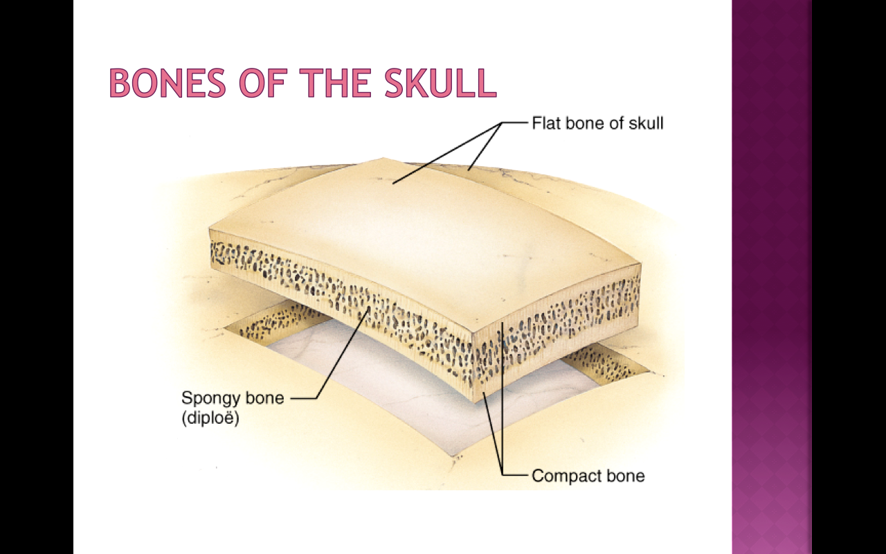
Table 1 describes the bone markings, which are illustrated in (figure 4). Cross section of long bone. The compact bone is made up of osteon. The cell line involved in osteogenesis consists of preosteoblasts, osteoblasts, osteocytes and bone. Bone is a dynamic biological tissue, composed of various metabolically active cells that are integrated into a rigid framework.
In A Cross Section Of A Bone You Can Usually See Two Types Of Bone Tissues What Are These Called Socratic from useruploads.socratic.org Vector illustration scheme of bone cross section. This slide contained a cross section of a very small bone, and you are looking at the entire thickness of the shaft of the bone. Sections of bone marrow tissue. Bone matrix and cells bone matrix osseous tissue is a connective tissue and like all connective tissues contains relatively few cells and large amounts of extracellular matrix. Compact bone, spongy bone, and bone marrow. Muscles and bones of the human body 12 photos of the muscles and bones of the human body anatomy bones of the human body quiz, major muscles and bones in the human body, muscles and bones in the human body, number of muscles and bones in the human body. Browse 4,253 bone cross section stock photos and images available, or search for human bone cross section to find more great stock photos and pictures. Beautiful tooth cross section illustration, deep blue background and sparkling lights around.
Sections of bone marrow tissue.
Explaned distal and proximal epiphysis. The large dark spots are passages for blood vessels and nerves. This is known as the periosteum. Internal structure of a human long bone, with a magnified cross section of the interior. Section of bone marrow affected by myeloma seen under a microscope. Smartdraw includes 1000s of professional healthcare and anatomy chart templates that you can modify and make your own. Two types of bone tissues in cross section of a long bone : Would it be a good thing to show the epiphyseal plate? The compact bone is made up of osteon. The cell line involved in osteogenesis consists of preosteoblasts, osteoblasts, osteocytes and bone. I don't find it enhances the image. The cortical bone equivalent area of the cross‐section of the region of interest (femoral neck or shaft), with all soft tissue voids (trabecular and cellular spaces) eliminated (cm 2). 100x first focus in the compact decalcified bone (cb) on the left part of the image, you can see small dots, which are.
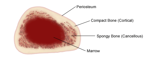
The central tubular region of the bone, called the diaphysis, flares outward near the end to form the metaphysis, which contains a largely cancellous, or spongy, interior. Cross section of long bone. Browse 53 bone marrow cross section stock photos and images available, or search for bone cross section or bone cells to find more great stock photos and pictures. The large dark spots are passages for blood vessels and nerves. Terms in this set (3) epiphysis.
Schematic Diagram Of Long Bone Cross Section 47 Download Scientific Diagram from www.researchgate.net The cortical bone equivalent area of the cross‐section of the region of interest (femoral neck or shaft), with all soft tissue voids (trabecular and cellular spaces) eliminated (cm 2). End of a long bone. The compact bone is made up of osteon. The surface features of bones vary considerably, depending on the function and location in the body. Cross section of long bone. Vector illustration scheme of bone cross section. Marrow in the shaft of long bones is typically yellow, with red marrow in the head through the cancellous bone. This is known as the periosteum.
End of a long bone.
While it is not as hard as compact bone, spongy bone plays an important role of protecting the marrow where blood cells are produced. Bone markings the surface features of bones vary considerably, depending on the function and location in the body. There are trabeculae in spongy bone which gives its sponge like appearance. Bone matrix and cells bone matrix osseous tissue is a connective tissue and like all connective tissues contains relatively few cells and large amounts of extracellular matrix. The upper (biting) surfaces of the tooth are at top, with the lower sections (bottom) embedded in the gums and jaw bone (not shown). Explaned distal and proximal epiphysis. The cell line involved in osteogenesis consists of preosteoblasts, osteoblasts, osteocytes and bone. Start studying cross section of bone. As the names suggest compact bone looks compact and the spongy bone looks like sponges. An outer 'fibrous layer' containing mainly fibroblasts, and an inner 'cambium layer' containing progenitor cells. 100x first focus in the compact decalcified bone (cb) on the left part of the image, you can see small dots, which are. Diagram with articular cartilage, marrow, spongy bone, medullary cavity, endosteum, diaphysis, and periosteum. Cross section of mandible at first molar region showing cortical and spongy bone basic concepts in osteogenesis.
Beautiful tooth cross section illustration, deep blue background and sparkling lights around. End of a long bone. Compact bone is the outer layer and the spongy bone forms the inner layer. Start studying cross section of bone. Section of bone marrow affected by myeloma seen under a microscope.
7 Inside Bones Laptops 60 Minutes What S Going On In Mr Solarz Class from psolarz.weebly.com Browse 53 bone marrow cross section stock photos and images available, or search for bone cross section or bone cells to find more great stock photos and pictures. Bone matrix and cells bone matrix osseous tissue is a connective tissue and like all connective tissues contains relatively few cells and large amounts of extracellular matrix. Cross section of long bone. To the left is muscle tissue, and to the right is bone marrow. Diagram with articular cartilage, marrow, spongy bone, medullary cavity, endosteum, diaphysis, and periosteum. The large dark spots are passages for blood vessels and nerves. Section of bone marrow affected by myeloma seen under a microscope. An outer 'fibrous layer' containing mainly fibroblasts, and an inner 'cambium layer' containing progenitor cells.
Cross section of long bone.
Explaned distal and proximal epiphysis. Two types of bone tissues in cross section of a long bone : Vector illustration scheme of bone cross section. Bone · february 15, 2021. Cross section of mandible at first molar region showing cortical and spongy bone basic concepts in osteogenesis. And why does the marrow stop where it does, and so sharply? Table 1 describes the bone markings, which are illustrated in (figure 4). They are obtained by taking imaginary slices perpendicular to the main axis of organs, vessels, nerves, bones, soft tissue, or even the entire human body. The central tubular region of the bone, called the diaphysis, flares outward near the end to form the metaphysis, which contains a largely cancellous, or spongy, interior. Marrow in the shaft of long bones is typically yellow, with red marrow in the head through the cancellous bone. The large dark spots are passages for blood vessels and nerves. Cross‐sectional area is derived from the integral of the bone mass profile across the narrow region. Browse 53 bone marrow cross section stock photos and images available, or search for bone cross section or bone cells to find more great stock photos and pictures.

0 Comments:
Posting Komentar